

Goat Anti-Rabbit IgG antibody (DyLight594)
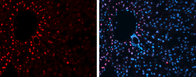
Immunofluorescence photomicrographs of frozen sections of mouse liver.
Red: Histone H3K27me3 (trimethyl Lys27) antibody (GTX121184) diluted at 1:200. The signal was developed using goat anti-rabbit IgG antibody (Dylight594) (GTX213110-05).
Blue: Nuclear staining with Hoechst 33342.
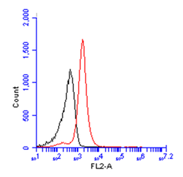
BrdU antibody (GTX128091) detects BrdU protein from HeLa treated with 10 μM BrdU for 30 minutes and fixed in 4% paraformaldehyde at 4ºC for 15 minutes by flow cytometry analysis.
Black: Unlabelled sample was used as a control.
Red: BrdU antibody (GTX128091) dilution: 1:50.
The Rabbit IgG antibody (DyLight594) (GTX213110-05) was used to detect the primary antibody.
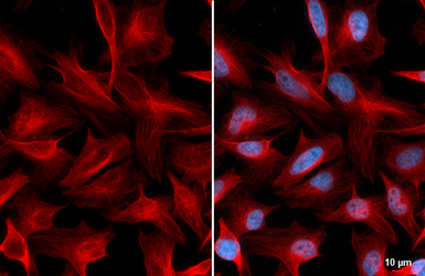
beta Tubulin antibody detects beta Tubulin protein at cytoskeleton by immunofluorescent analysis. Sample: HeLa cells were fixed in 4% paraformaldehyde at RT for 15 min. Red: beta Tubulin stained by beta Tubulin antibody (GTX101279) diluted at 1:100. The signal was developed using Goat Anti-Rabbit IgG antibody (DyLight594) (GTX213110-05) diluted at 1:500. Blue: Fluoroshield with DAPI (GTX30920).