

Goat Anti-Rabbit IgG antibody, pre-adsorbed (HRP)
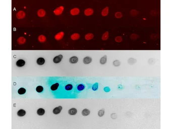
Dot blot analysis of serially diluted rabbit IgG using GTX27090 Goat Anti-Rabbit IgG antibody, pre-adsorbed (HRP) detected by different platforms.
Dilution : 1:20000
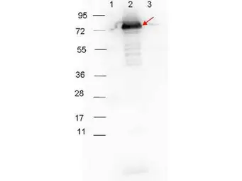
WB analysis of various samples using anti-p27 antibody followed by GTX27090 Goat Anti-Rabbit IgG antibody, pre-adsorbed (HRP).
Lane 1 : Protein ladder
Lane 2 : MBP-p27 protein
Lane 3 : MBP
Dilution : 1:40000
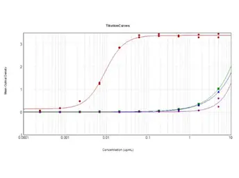
ELISA analysis of various samples using serially diluted GTX27090 Goat Anti-Rabbit IgG antibody, pre-adsorbed (HRP).
Red : Rabbit IgG
Green : Bovine IgG
Blue : Chicken IgG
Purple : Goat IgG
Coating : 10 μg

GeneTex secondary antibodies detect rabbit primary antibodies in a variety of platforms. Shown here is a serial 1:1 dilution of control rabbit IgG protein 250ng starting total load Co-incubated with GeneTex HRP conjugated Goat anti Rabbit IgG antibody (GTX27090) and Dylight 649 conjugated goat anti Rabbit IgG antiody at 1:20K in blocking buffer.
GTX27090 secondary antibody was used at 1:40,000 in blocking buffer to detect a rabbit primary antibody by Western Blot. Anti p27 antibody (GTX48799, 1:1000 RT 30 minutes) showed detection of 0.1 μg of recombinant p27 protein. Lane 1: Molecular weight markers. Lane 2: MBP-p27 fusion protein (arrow; expected MW: 73.3 kDa). Lane 3: MBP alone. Protein was run on a 4-20% gel, then transferred to 0.45 μm nitrocellulose and blocked with 1% BSA-TTBS overnight at 4ºC. Blot was imaged on the VersaDoc MP 4000 imaging system (Bio-Rad).