- Specifications
Product Description
Intracellular Total ROS Activity Assay Kit (Orange Fluorescence) is used for the measurement of intracellular total ROS activity.
Suitable Sample
Live Cells
Excitation/<br>Emission (nm)
540/570 nm
Detection Method
Fluorometric
Instrument Platform
Flow Cytometer, Fluorescence Microplate Reader, Fluorescence Microscope
Regulation Status
For research use only (RUO)
Storage Instruction
Store the kit in desiccated environment at -20°C and avoid from light.
Note
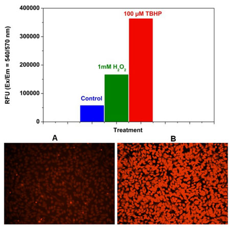
Detection of ROS in Hela cells.
- Applications
Quantification
- Publication Reference
- Electron Redistribution in Iridium-Iron Dual-Metal-Atom Active Sites Enables Synergistic Enhancement for H2O2 Decomposition.
Zhiwei Wang, Lu Peng, Ping Zhu, Wenlong Wang, Cheng Yang, Hong-Ying Hu, Qianyuan Wu.
ACS Nano 2024 Jan; 18(4):2885.
Application:Quant, Hamster, CHO-K1 cell line.
- Vacuum ultraviolet irradiation for reduction of the toxicity of wastewater towards mammalian cells: Removal mechanism, changes in organic compounds, and toxicity alternatives.
Liu He, Wen-Long Wang, De-Xiu Wu, Shao-Yu Wang, Xiao Xiao, He-Qing Zhang, Min-Yong Lee, Qian-Yuan Wu.
Environment International 2023 Dec; 182:108314.
Application:Quant, Hamster, CHO-K1 cells.
- Overlooked Cytotoxicity and Genotoxicity to Mammalian Cells Caused by the Oxidant Peroxymonosulfate during Wastewater Treatment Compared with the Sulfate Radical-Based Ultraviolet/Peroxymonosulfate Process.
Ye Du, Wen-Long Wang, Zhi-Wei Wang, Chang-Jie Yuan, Ming-Qi Ye, Qian-Yuan Wu.
Environmental Science & Technology 2023 Feb; 57(8):3311.
Application:Quant, Hamster, CHO-k1 cells.
- Multi-endpoint assays reveal more severe toxicity induced by chloraminated effluent organic matter than chloraminated natural organic matter.
Hai-Yan Wang, De-Xiu Wu, Ye Du, Xiao-Tong Lv, Qian-Yuan Wu.
Journal of Environmental Sciences 2024 Jan; 135:310.
Application:Func, Hamster, CHO cells.
- Reduction of cytotoxicity and DNA double-strand break effects of wastewater by ferrate(VI): Roles of oxidation and coagulation.
Qian-Yuan Wu, Xue-Si Lu, Ming-Bao Feng, Wen-Long Wang, Ye Du, Lu-Lin Yang, Hong-Ying Hu.
Water Research 2021 Oct; 205:117667.
Application:Func, Hamster, CHO cells.
- Toxicity of Ozonated Wastewater to HepG2 Cells: Taking Full Account of Nonvolatile, Volatile, and Inorganic Byproducts.
Qian-Yuan Wu, Lu-Lin Yang, Ye Du, Zi-Fan Liang, Wen-Long Wang, Zhi-Min Song, De-Xiu Wu.
Environmental Science & Technology 2021 Aug; 55(15):10597.
Application:Func, IF, Human, HepG2 cells.
- Non-volatile disinfection byproducts are far more toxic to mammalian cells than volatile byproducts.
Qian-Yuan Wu, Zi-Fan Liang, Wen-Long Wang, Ye Du, Hong-Ying Hu, Lu-Lin Yang, Wen-Cheng Huang.
Water Research 2020 Sep; 183:116080.
Application:Func, IF, Quant, Hamster, CHO cells.
- Chlorinated effluent organic matter causes higher toxicity than chlorinated natural organic matter by inducing more intracellular reactive oxygen species.
Du Y, Wang WL, He T, Sun YX, Lv XT, Wu QY, Hu HY.
The Science of the Total Environment 2020 Jan; 701:134881.
Application:Func, Mouse, CHO cells.
- Study on the effect of LncRNA AK094457 on OX-LDL induced vascular smooth muscle cells.
Liu M, Song Y, Han Z.
American Journal of Translational Research 2019 Sep; 11(9):5623.
Application:Func, Human, Human vascular smooth muscle cells.
- Underestimated risk from ozonation of wastewater containing bromide: Both organic byproducts and bromate contributed to the toxicity increase.
Wu QY, Zhou YT, Li W, Zhang X, Du Y, Hu HY.
Water Research 2019 Oct; 162:43.
Application:Func, Mouse, CHO cells.
- Exposure to solar light reduces cytotoxicity of sewage effluents to mammalian cells: Roles of reactive oxygen and nitrogen species.
Du Y, Wu QY, Lv XT, Wang QP, Lu Y, Hu HY.
Water Research 2018 Jul; 143:570.
Application:Func, Mouse, CHO cells.
- Electron Redistribution in Iridium-Iron Dual-Metal-Atom Active Sites Enables Synergistic Enhancement for H2O2 Decomposition.
Intracellular Total ROS Activity Assay Kit (Orange Fluorescence)
Referencia KA4075
embalaje : 1Kit
Marca : Abnova
Images


