

Polyclonal Antibody to Ubiquitin C-terminal hydrolase 1, UCHL1
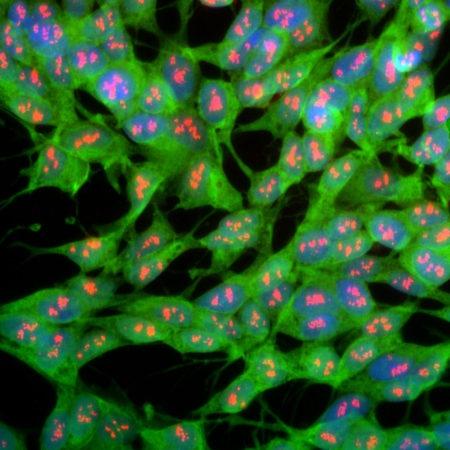
Figure-1: Immunofluorescent analysis of SH-SY5Y cells stained with rabbit pAb to UCHL1,(34-1125), dilution 1:1,000 in green, and costained with mouse mAb to fibrillarin,(34-1031), dilution 1:1,000 in red. The blue is Hoechst staining of nuclear DNA. The UCHL1 antibody produces strong staining of the cellular cytoplasm of these cells which share many properties with neurons, while the (34-1031) thumbnails_wrapper_product">


